The research focuses on microtubules, tiny structures within cells that ensure chromosomes are properly aligned before the cell divides. In cancer cells, these structures often move too quickly, causing errors in chromosome distribution that can contribute to the disease’s progression.
The team, which includes Dr. Patrik Risteski, Dr. Jelena Martinčić, Mihaela Jagrić, Ana Petelinec, and undergraduate student Erna Tintor, discovered the crucial role of microtubule movement in the accurate alignment and division of chromosomes. By adjusting the speed of microtubule movement, they were able to correct distribution errors in both healthy and cancer cells, providing insights into a mechanism that could potentially help in eliminating cells that show chromosome instability, a major driver of cancer.
Why Chromosome Alignment Is Crucial
Cancer cells often exhibit errors during division, known as chromosomal instability, which results in new cells having an incorrect number of chromosomes. This imbalance can affect their growth and behaviour, potentially enabling the cancer cells to develop resistance to treatments. However, targeting this chromosomal instability could serve as a strategy for new therapeutic interventions.
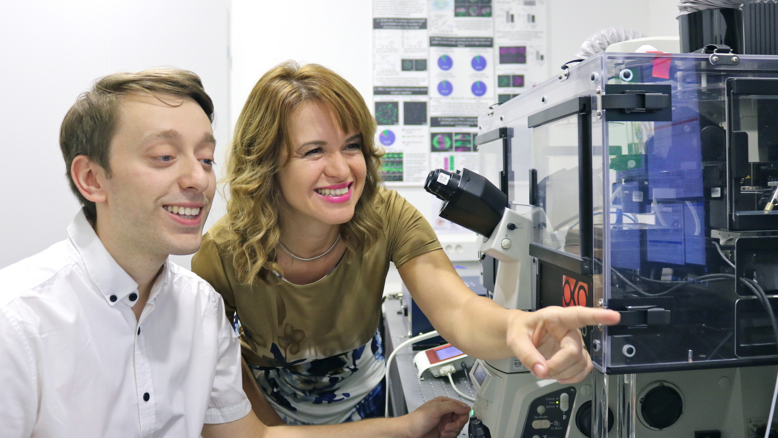
Mihaela Jagrić, Jelena Martinčić i Patrik Risteski
The study explored the possibility that controlling the speed of microtubules could correct these division errors. The team focused on specific areas where microtubules overlap and are propelled by motor proteins. By minimizing these overlaps, they managed to reduce the excessive microtubule activity and restore balance to the cellular division process.
Future Potential
The research utilized speckle microscopy, a method developed by Dr. Patrik Risteski during his Ph.D., which allows for the visualization of microtubules in a speckled pattern at extremely low concentrations. This innovative technique offers a detailed look at the dynamics of individual microtubules during cell division.
This study not only emphasizes the importance of microtubule movement in cell division but also the potential for future therapies targeting specific characteristics of cancer cells, such as misaligned chromosomes.
Support and Funding
This research was completely conducted at RBI, with funding from the European Research Council (ERC) Synergy Grant and the Croatian Science Foundation (HrZZ). Doctoral students Mihaela Jagrić and Ana Petelinec also received support from HrZZ. The development of the microscopy protocols occurred on equipment funded by the project "Innovative Microscopy Protocols for Interdisciplinary Research in Medicine," supported by the European Fund for Regional Development under the Operational Programme Competitiveness and Cohesion 2014-2020.


